Loculated Pleural Effusion X Ray : Pleural Effusion Springerlink - Method to facilitate drainage of loculated hemorrhagic or fibrinous nonhemorrhagic pleural fluid collections.
Loculated Pleural Effusion X Ray : Pleural Effusion Springerlink - Method to facilitate drainage of loculated hemorrhagic or fibrinous nonhemorrhagic pleural fluid collections.. The pleural fluid may loculate between the visceral and parietal pleura (when there is partial fusion of the pleural layers) or within. Numerous septic emboli have caused foci of infection in his lungs with associated pleural effusions and he has an associated characteristic vasculitis. This case highlights the atypical but unique presentation of a transudative pleural effusion and demonstrates the risk of repeated. Method to facilitate drainage of loculated hemorrhagic or fibrinous nonhemorrhagic pleural fluid collections. Pleural effusions can also form when there is transport of peritoneal fluid from the abdominal cavity through the diaphragm or via lymphatics from a subdiaphragmatic process.
Lateral decubitus films may show loculated pleural effusions assist the patient with relaxation measures to reduce oxygen demand; The lungs and the chest cavity both have a lining that consists of pleura, which is a thin membrane. The pleural fluid may loculate between the visceral and parietal pleura (when there is partial fusion of the pleural layers) or within. Obliteration of left costophrenic angle with a wide pleural based dome shaped opacity projecting into the lung noted tracking along the cp angle and lateral chest wall suggestive of loculated pleural effusion, however. Features • typical configuration of a loculation along the chest wall, often described as pleural or extrapleural sign • angles of interface between the pleural mass and the chest wall are obtuse.
Radiographic Examinations Thoracic Key from thoracickey.com The patient's history and physical exam may indicate a presumptive. Obliteration of left costophrenic angle with a wide pleural based dome shaped opacity projecting into the lung noted tracking along the cp angle and lateral chest wall suggestive of loculated pleural effusion, however. Ct scan is the most sensitive modality for detection of presence of minimal fluid. This case highlights the atypical but unique presentation of a transudative pleural effusion and demonstrates the risk of repeated. Suspected parenchymal or pleural pathology. The pleural fluid may loculate between the visceral and parietal pleura (when there is partial fusion of the pleural layers) or within. Check for pleural thickening and pleural effusions. Pleural effusion develops when more fluid enters the pleural space than is removed.
In the usa approximately 1.5 million people are diagnosed with a pleural effusion each year 2.
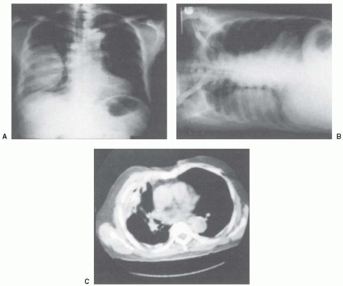
Loculated pleural effusion masquerading as mediastinal tumour had been reported but pleural effusion that conformed to the contour of a lung lobe is rare. Suspected parenchymal or pleural pathology. If you miss a tension pneumothorax you risk your patient's. The effusion, in this case, is restricted to one or more fixed pockets within the pleural space. Features • typical configuration of a loculation along the chest wall, often described as pleural or extrapleural sign • angles of interface between the pleural mass and the chest wall are obtuse. Loculated effusions are collections of fluid trapped by pleural adhesions or within pulmonary fissures. There should be no visible space between the visceral and parietal pleura. Concave meniscus (horizontal in case of. A pleural effusion is an abnormal collection of fluid within the pleural space. The annual incidence of pleural effusion in the developed world has been estimated at 320 per 100,000 population per year 1. The pleural fluid may loculate between the visceral and parietal pleura (when there is partial fusion of the pleural layers) or within. In the usa approximately 1.5 million people are diagnosed with a pleural effusion each year 2. Ct scans show more detail than.
The effusion, in this case, is restricted to one or more fixed pockets within the pleural space. Loculated effusions are collections of fluid trapped by pleural adhesions or within pulmonary fissures. Suspected parenchymal or pleural pathology. Lateral decubitus films may show loculated pleural effusions assist the patient with relaxation measures to reduce oxygen demand; In healthy lungs, these membranes ensure that a small amount of liquid is present between the lungs.
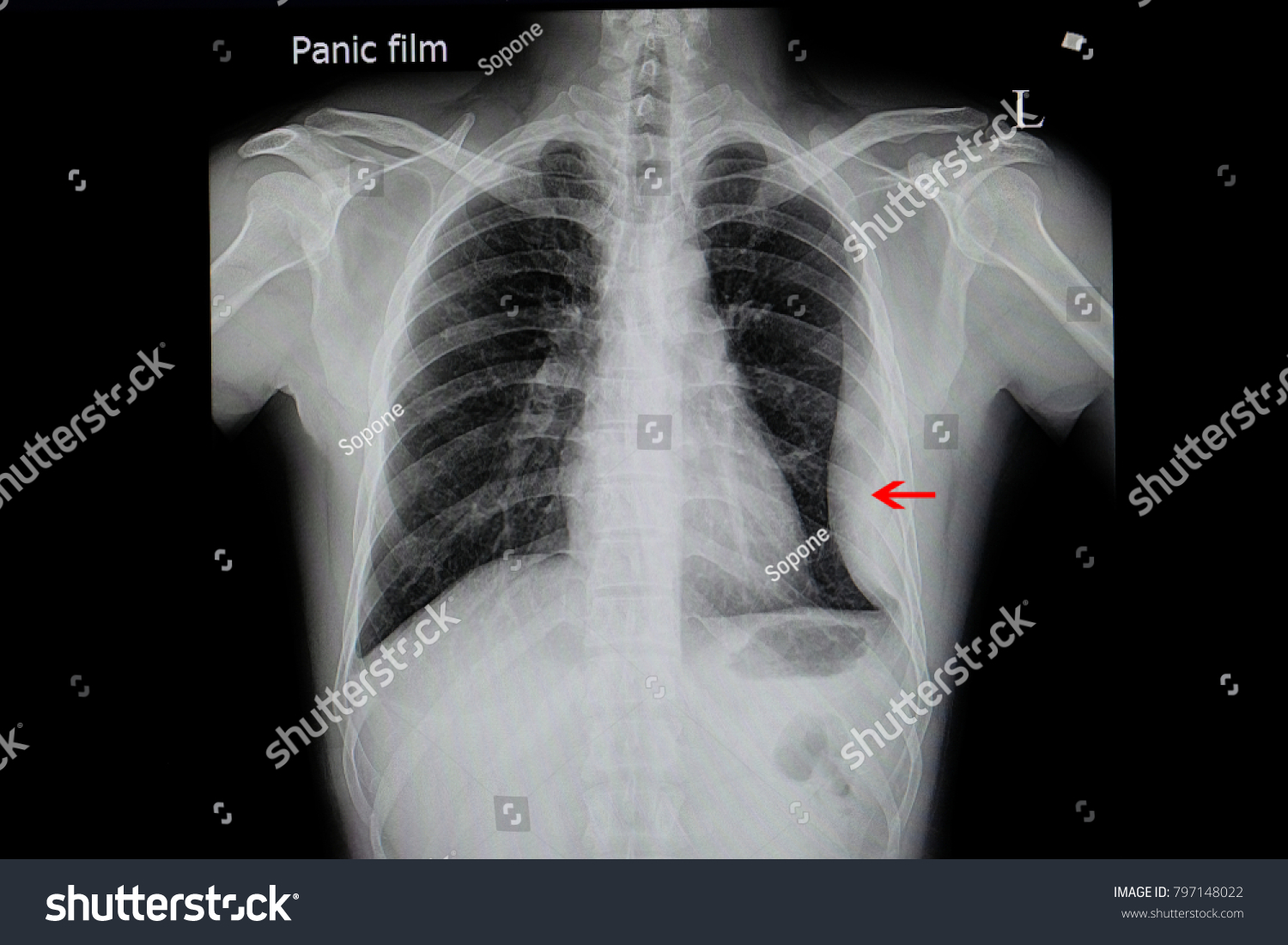
Chest Xray Film Patient Loculated Pleural Stock Photo Edit Now 797148022 from image.shutterstock.com In the usa approximately 1.5 million people are diagnosed with a pleural effusion each year 2. Pleura is a mesothelial lined sac that envelopes the lungs and comprises of 2 membranous walls i.e. The pleura and pleural spaces are only visible when abnormal. A pleural effusion is accumulation of excessive fluid in the pleural space, the potential space that surrounds each lung. Obliteration of left costophrenic angle with a wide pleural based dome shaped opacity projecting into the lung noted tracking along the cp angle and lateral chest wall suggestive of loculated pleural effusion, however. Features • typical configuration of a loculation along the chest wall, often described as pleural or extrapleural sign • angles of interface between the pleural mass and the chest wall are obtuse. The plain chest radiographic features of pleural effusion are usually characteristic. Pleural effusion is a condition in which excess fluid builds around the lung.
Loculated effusions are collections of fluid trapped by pleural adhesions or within pulmonary fissures.
Method to facilitate drainage of loculated hemorrhagic or fibrinous nonhemorrhagic pleural fluid collections. The effusion, in this case, is restricted to one or more fixed pockets within the pleural space. It allows distinction between free and loculated fluid showing its extent and localization. The plain chest radiographic features of pleural effusion are usually characteristic. Concave meniscus (horizontal in case of. More than one half of these massive pleural effusions are caused by malignancy; Ct scans show more detail than. The patient's history and physical exam may indicate a presumptive. Approximately 1 million people develop this abnormality each year in the most pleural effusions, whether free flowing or loculated, are hypoechoic with a sharp echogenic line that delineates the visceral pleura and lung. What procedures and tests diagnose pleural effusions? Pleural effusions can also form when there is transport of peritoneal fluid from the abdominal cavity through the diaphragm or via lymphatics from a subdiaphragmatic process. This case highlights the atypical but unique presentation of a transudative pleural effusion and demonstrates the risk of repeated. Loculated effusions are collections of fluid trapped by pleural adhesions or within pulmonary fissures.
Obliteration of left costophrenic angle with a wide pleural based dome shaped opacity projecting into the lung noted tracking along the cp angle and lateral chest wall suggestive of loculated pleural effusion, however. More than one half of these massive pleural effusions are caused by malignancy; A pleural effusion is accumulation of excessive fluid in the pleural space, the potential space that surrounds each lung. no change in position of effusion withchange in position of chest. Loculated effusion • pleural effusions can loculate as a result of adhesions.
Malignant Pleural Effusion Pulmonology Advisor from www.pulmonologyadvisor.com A parasternal long axis and subcostal views are shown. Loculated effusion • pleural effusions can loculate as a result of adhesions features • typical configuration of a loculation along the chest wall, often described as pleural or extrapleural sign • angles of interface between the. In the usa approximately 1.5 million people are diagnosed with a pleural effusion each year 2. If you miss a tension pneumothorax you risk your patient's. The pleura and pleural spaces are only visible when abnormal. .or fibrinous nonhemorrhagic loculated pleural collections in 11 patients with 13 loculated pleural collections. Role model positive coping strategies. It allows distinction between free and loculated fluid showing its extent and localization.
Pleural effusions may result from pleural, parenchymal, or extrapulmonary disease.
Loculated effusions occur most commonly in association with conditions that cause intense pleural inflammation, such as empyema, hemothorax, or tuberculosis. Suspected parenchymal or pleural pathology. Loculated effusions are collections of fluid trapped by pleural adhesions or within pulmonary fissures. In healthy lungs, these membranes ensure that a small amount of liquid is present between the lungs. Ct scan is the most sensitive modality for detection of presence of minimal fluid. Ct scans show more detail than. Loculated pleural effusion masquerading as mediastinal tumour had been reported but pleural effusion that conformed to the contour of a lung lobe is rare. Other causes are complicated parapneumonic effusion. Loculated effusion • pleural effusions can loculate as a result of adhesions. Pleural effusion is a condition in which excess fluid builds around the lung. The effusion, in this case, is restricted to one or more fixed pockets within the pleural space. Pleural effusions can also form when there is transport of peritoneal fluid from the abdominal cavity through the diaphragm or via lymphatics from a subdiaphragmatic process. Role model positive coping strategies.
Ct scan is the most sensitive modality for detection of presence of minimal fluid loculated pleural effusion. Concave meniscus (horizontal in case of.


0 Komentar